


A nanoscale project represents a giant leap forward in understanding the human brain.
With more than 1.4 petabytes of electron microscopy imaging data, a team of scientists has reconstructed a teeny-tiny cubic segment of the human brain.
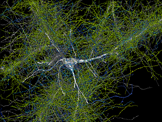
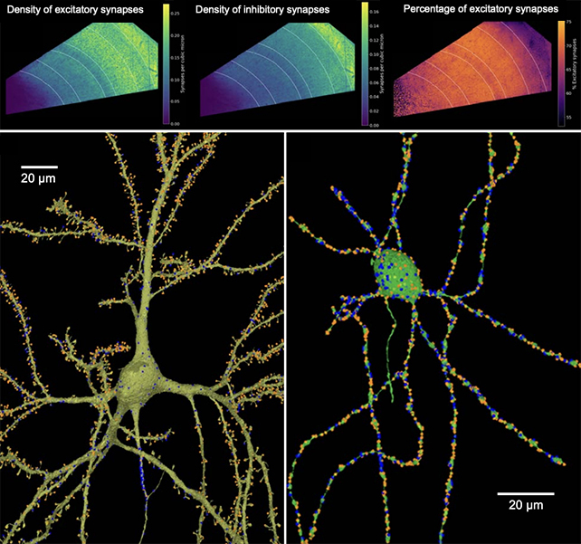
It’s just a millimeter on each side – but 57,000 cells, 150 million synapses, and 230 millimeters of ultrafine veins are all packed into that microscopic space.
The work of almost a decade, it’s the largest and most detailed reproduction of the human brain to date down to the resolution of the synapses, the structures that allow neurons to transmit signals between them.
“The word ‘fragment’ is ironic,” says neuroscientist Jeff Lichtman of Harvard University. “A terabyte is, for most people, gigantic, yet a fragment of a human brain – just a miniscule, teeny-weeny little bit of human brain – is still thousands of terabytes.”
The human brain is notoriously complex. Across the animal kingdom, the functions performed by most of the vital organs are more or less the same, but the human brain is in a league of its own.
It’s also very difficult to study; there’s so much going on in there, on such miniscule scales, that we’ve been unable to understand the synaptic circuitry in detail.
Each human brain contains billions of neurons, firing signals back and forth via trillions of synapses, the command center from which the human body is run.
A deeper understanding of the way this dazzlingly complicated organ operates would confer profound benefits to our studies of brain function and disorders, from injury to mental illness to dementia.
To that end, Lichtman and colleagues have been working on what they call a “connectome” – a map of the brain and all its wiring that could help better understand when that wiring is askew.
The current goal for the connectomics project is the reproduction of an entire mouse brain, but using similar techniques to reconstruct at least segments of the human brain can only advance our knowledge faster.
The team’s reconstruction was based on a sample of human brain excised from an epilepsy patient during surgery to access an underlying lesion. The sample was fixed, stained with heavy metals to accentuate the details, embedded in resin, and sectioned into 5,019 slices, with a mean thickness of 33.9 nanometers, collected on tape.
The researchers used high-throughput serial section electron microscopy to image this tiny piece of tissue in mind-numbing detail, generating 1.4 petabytes (1,400 terabytes) of data.
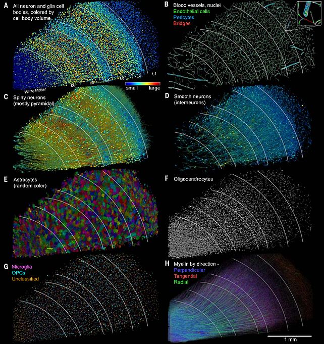
This data was analyzed with specially developed techniques and algorithms, generating, the researchers say, “a 3D reconstruction of nearly every cell and process in the aligned volume.”
This reconstruction, named H01, has already revealed some previously unseen fine details about the human brain. The team was surprised to note that glia, or non-neuronal cells, outnumbered neurons 2:1 in the sample, and the most common cell type was oligodendrocytes – cells that help coat axons in protective myelin.
Each neuron had thousands of relatively weak connections, but the researchers found rare, powerful sets of axons connected by 50 synapses. And they found that a small number of axons are arranged in unusual, extensive whorls.
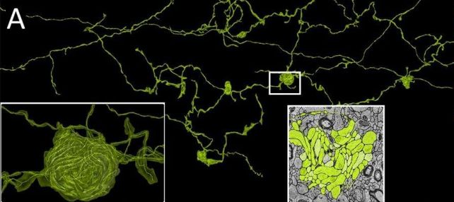
Because the sample was taken from a patient with epilepsy, it’s unclear whether these are normal, but rare, features of the human brain, or linked to the patient’s disorder. Either way, though, the work has revealed the vast breadth and depth of the chasm of our understanding of the brain.
The next step in the team’s work involves trying to understand the formation of the mouse hippocampus, a brain region heavily involved in learning and memory.
“If we get to a point where doing a whole mouse brain becomes routine, you could think about doing it in say, animal models of autism,” Lichtman explained last year to The Harvard Gazette.
“There is this level of understanding about brains that presently doesn’t exist. We know about the outward manifestations of behavior. We know about some of the molecules that are perturbed. But in between the wiring diagrams, until now, there was no way to see them. Now, there is a way.”
The research has been published in Science, and the data and reconstruction of H01 have been made freely available on a dedicated website.
Reference :
Reference link


+ There are no comments
Add yours